Development of Deep Learning Models to Reduce Artifacts in CBCT Images
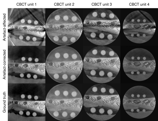
Cone-beam computed tomography (CBCT) is a widely used 3D imaging modality, particularly in implant dentistry, orthopedics, and image-guided radiation therapy. While it offers several advantages over conventional CT, CBCT is highly susceptible to artifacts caused by high-density objects such as dental implants. These artifacts degrade image quality, hinder accurate analysis, and potentially compromise diagnostic tasks.
Using smaller fields of view (FOV) improves image sharpness and reduces patient radiation exposure. However, currently used metal artifact reduction algorithms are less effective when dental implants are not visible in the reconstructed images - an issue that can arise with small FOVs.
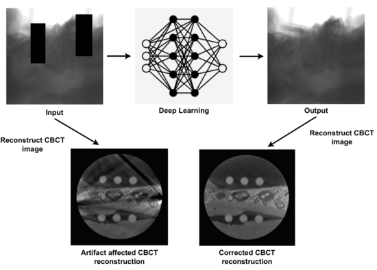
The medical field has shown a growing interest in deep learning methods over the past decade. Several deep learning models have been proposed for artifact reduction in CT/CBCT images. Many of these approaches are limited to 2D images and do not account for cases where implants are located both within and outside the exomass.
The goal of this project is to develop 3D deep learning models capable of removing artifacts caused by dental implants, regardless of their visibility in the reconstructed images, that could be integrated into CBCT scanner software. The data for this project was provided by the University Center of Dental Medicine Basel (UBZ).