

SA-Profile: Automated Sulcus Angle Profiling from Super-Resolution MRI
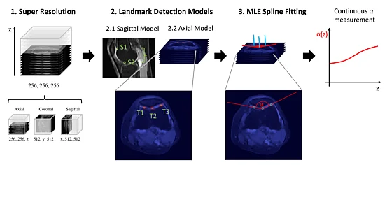
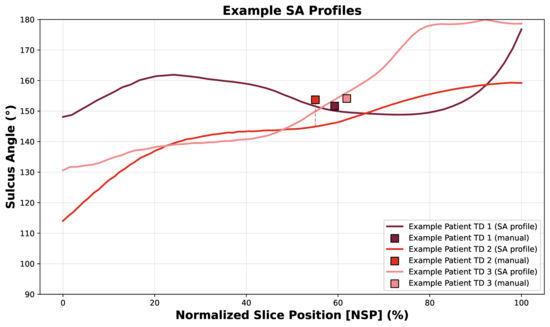
The sulcus angle is commonly used to assess trochlear morphology but is typically measured on a single axial MR slice. In this project, we developed an automated framework that extends this conventional measurement to a continuous sulcus angle profile across the trochlear region. Clinical axial, coronal, and sagittal MR scans are combined using implicit neural representations to reconstruct a high-resolution volume, from which sulcus angles are automatically determined using landmark detection networks. The method was evaluated on the public fastMRI dataset and an in-house cohort of patients with trochlear dysplasia, demonstrating distinct mean sulcus angle profiles between the two cohorts. The approach enables a more comprehensive description of trochlear morphology without requiring additional imaging and remains directly linked to established clinical assessment.
For more information about this project, please contact Michael Wehrli.

Michael Wehrli

Lorenzo Pettinari

Leo Widmer

Sidaty El Hadramy

Paul Friedrich

Philippe C. Cattin
- Luana Brahaj, Department of Biomedical Engineering, University of Basel
- Dr. med Edwin Li, Olten Cantonal Hospital
- Prof. Dr. Carol-Claudius Hasler, Department of Orthopedics, University Children's Hospital Basel
Werner Siemens Foundation through the MIRACLE II project