This site requires JavaScript to work properly.
How do I activate JavaScript in this web browser?
Event Registration
Documents
Search
Search
Search
Department of
Biomedical Engineering
Search
Search
Search
News
Events
Newsletter
Event Online Registration
Education
Master of Science
PhD in Biomedical Engineering
Research
Innovation
Collaboration
Patents
Spin-Offs
Services
Public
Events
Material about the DBE
Visit Us
About us
People
Organisation
Contact and Directions
Culture
Career
History of the DBE
Policies & Guidelines
First Encounter
Alumni
Website Root
Research
Core Facilities
Core Facility MiNa
Equipment
nanotom® m
News
Events
Newsletter
Event Online Registration
Education
Master of Science
PhD in Biomedical Engineering
Research
Biomaterials Science and Regenerative Medicine
Clinical Biomechanics
Data Driven Modelling and Analysis
Emerging Analytical and Forensic Technologies
Lasers & Robotics
Medical Imaging
Flagship Project MIRACLE
Core Facilities
All Research Groups
Innovation
Collaboration
Patents
Spin-Offs
Services
Public
Events
Material about the DBE
Visit Us
About us
People
Organisation
Contact and Directions
Culture
Career
History of the DBE
Policies & Guidelines
First Encounter
Alumni
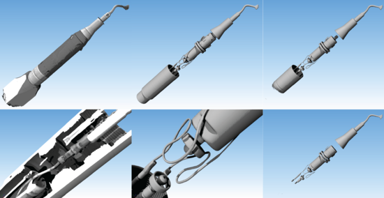
nanotom® m (phoenix|x-ray, now Baker Hughes)
Scanning parameters
maximal sample diameter: 240 mm
maximal sample height: 250 mm
maximal sample weight: 3 kg
smallest effective pixel size: 0.3 µm
reconstructed volume after single scan: 3052 × 3052 × 2400 voxels
reconstructed volume after 'off-axis' scan: 4624 × 4624 × 2400 voxels
typical scanning time (depending on application): 5 - 480 min
X-ray detector
temperature-stabilized high dynamic GE DXR
7 MP (3072 x 2400)
100 µm pixel size
Nanofocus X-ray source
spot size: < 1 µm
acceleration voltage: 40 - 180 kV
maximal power: 20 W
available targets: tungsten on CVD diamond & molybdenum
nanotom® m brochure
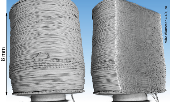
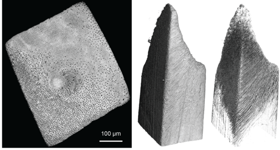
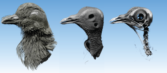
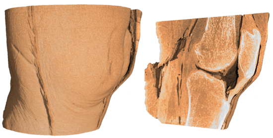
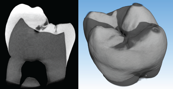
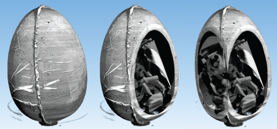
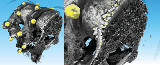
Examples acquired with nanotom® m
Quick Links
Social Media
Linkedin
Instagram
YouTube
© University of Basel
Privacy Policy
Disclaimer
To top