Technologies
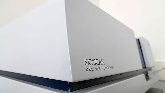
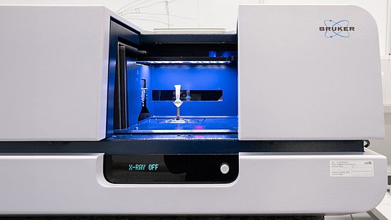
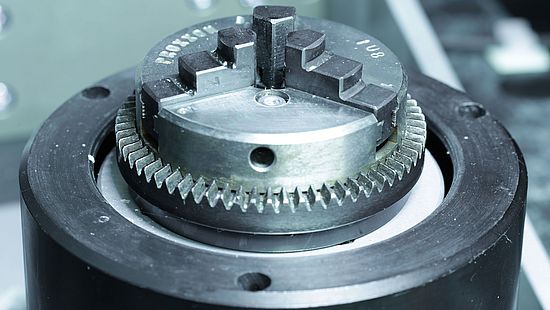
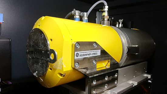
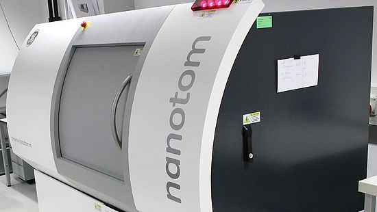
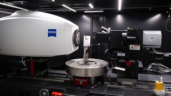
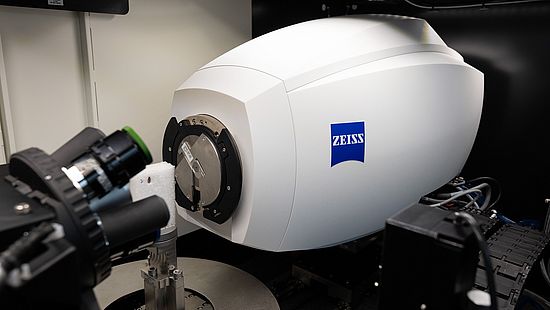
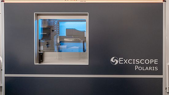
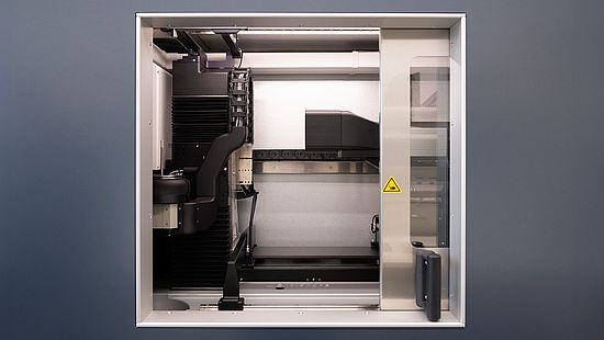
The Core Facility MiNa houses four microtomography systems. Two state-of-the-art systems, namely nanotom® m (phoenix|x-ray, Waygate Technologies, Baker Hughes, Wunstorf, Germany) and Skyscan 1275 (Bruker microCT, Kontich, Belgium) have been successfully used since 2012. Two next-generation devices funded by the SNSF R'Equip and the University of Basel were installed in fall 2023: xradia 610 Versa (Zeiss, Oberkochen, Germany) and Polaris (Excsicope, Kista, Sweden).
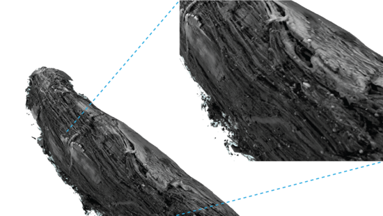
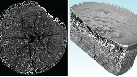
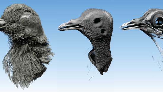
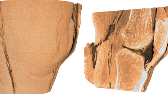
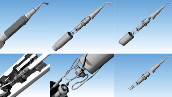
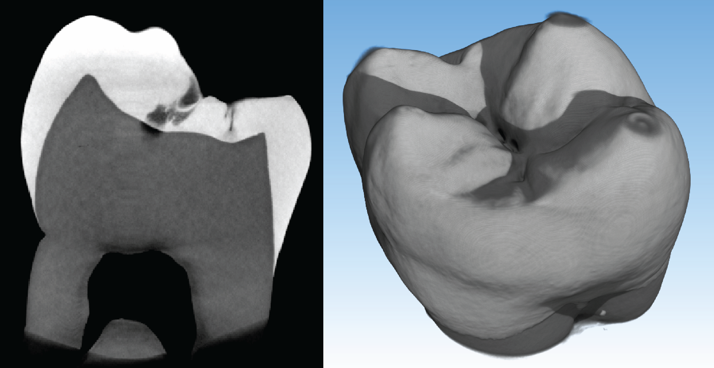
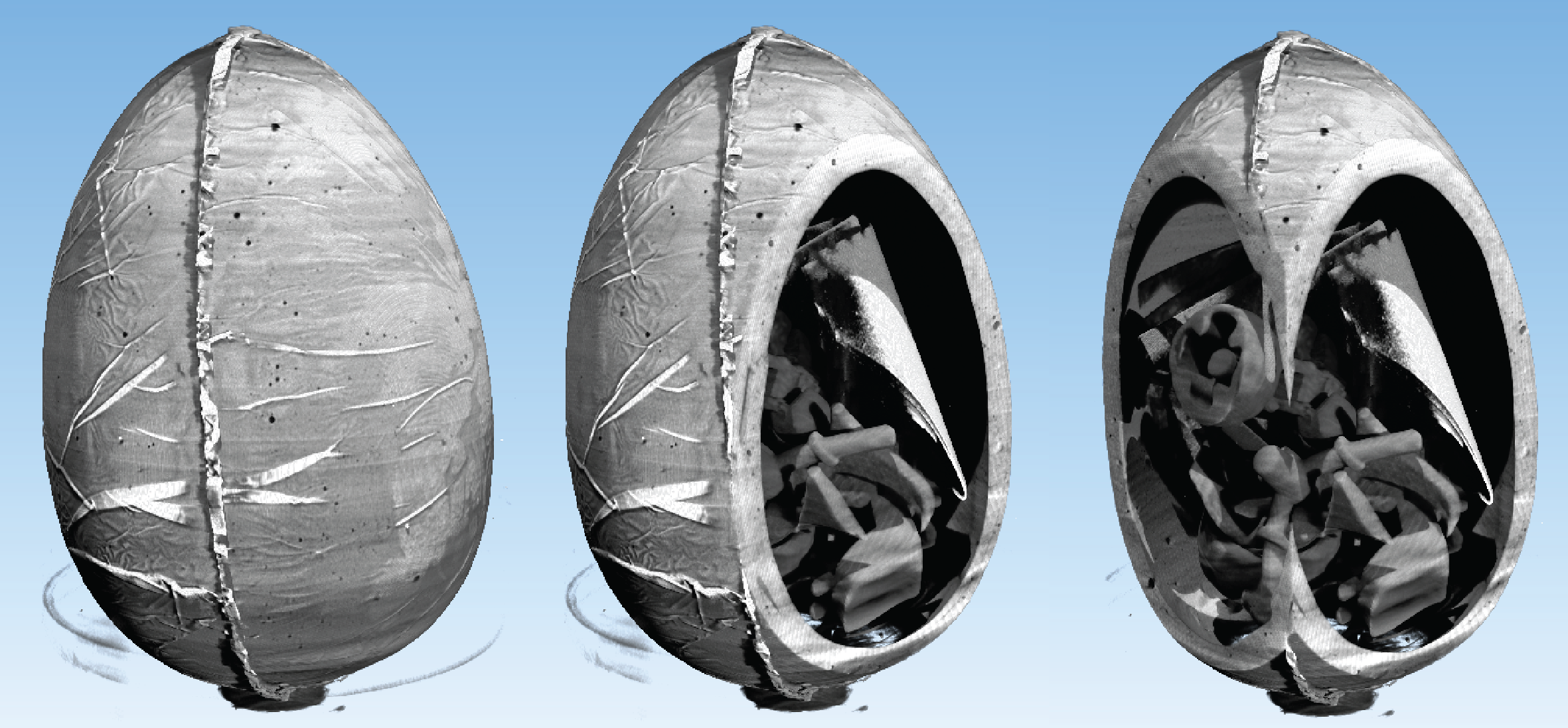
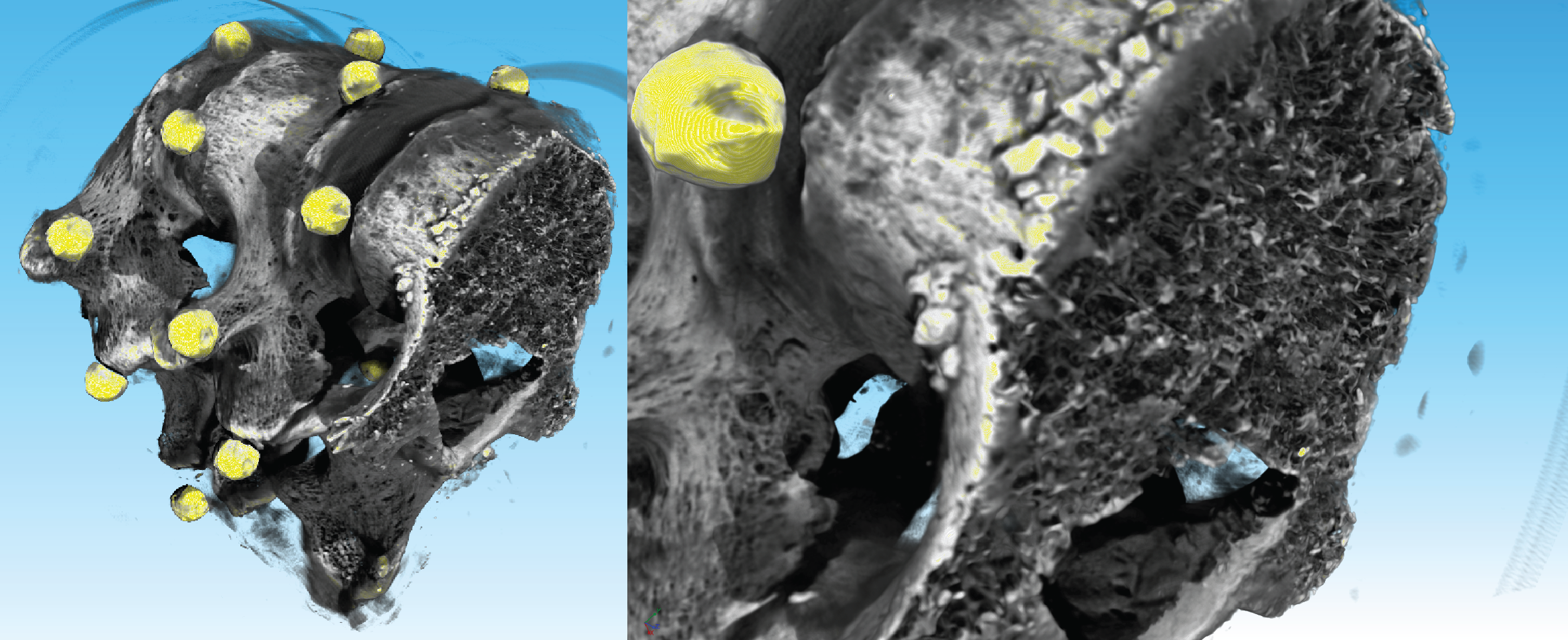
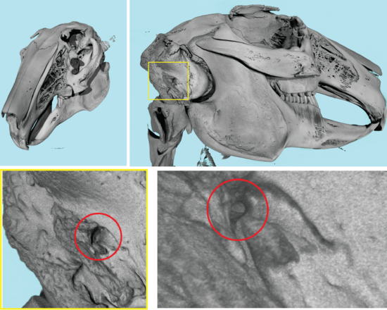
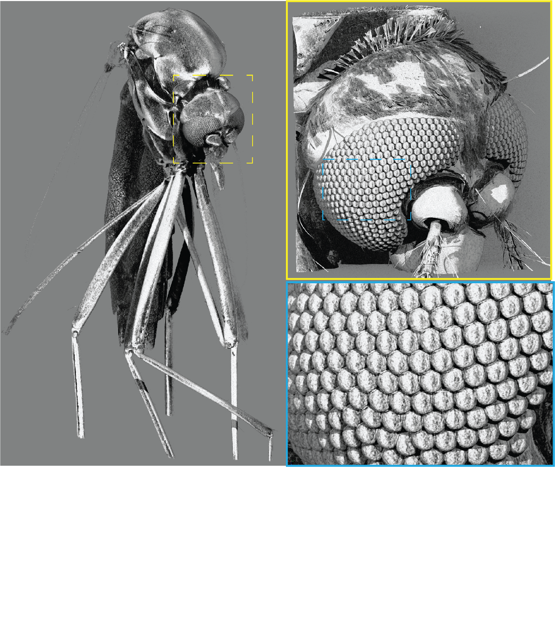
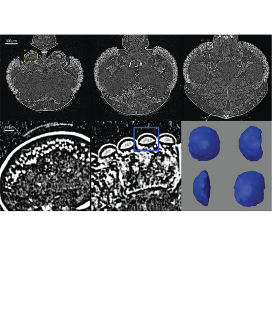
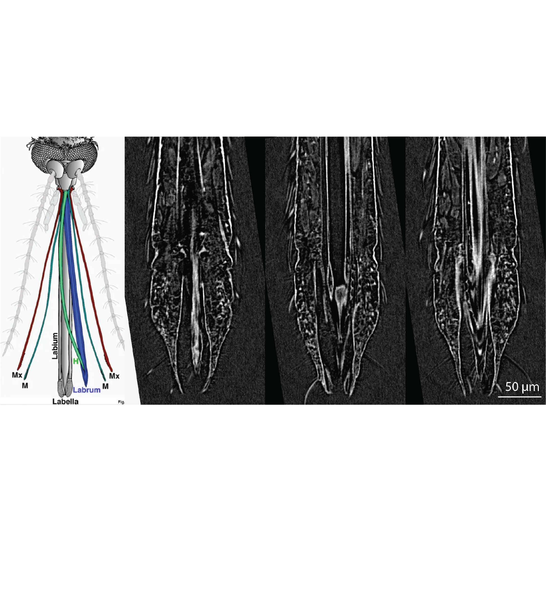
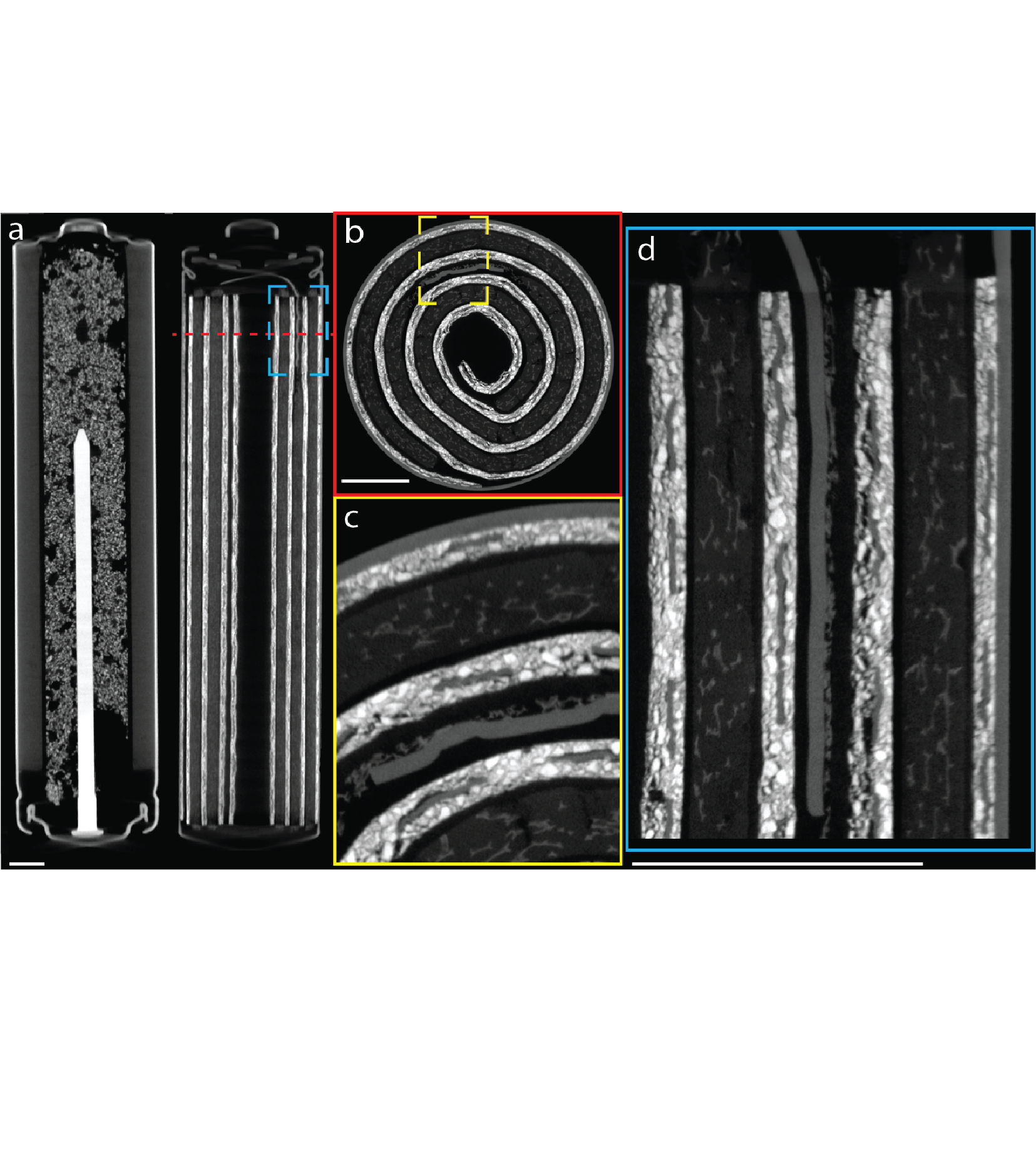
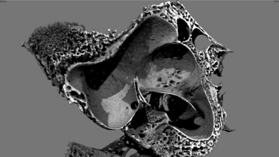
With these systems, the Core Facility can support a broad range of applications, including life sciences, pharmaceutical research, energy storage, electronics, medical implants, and the analysis of unique museum objects.
Interested in our services?
Contact us for more details or a price estimation of our services.
Scanning parameters
- maximal sample diameter: 96 mm
- maximal sample height: 120 mm
- smallest effective pixel size: 4 µm
- reconstructed volume after single scan: 1944 x 1944 x 1160 voxels
- typical scanning time (depending on application): 10 - 120 min
X-ray detector
- active pixel CMOS flat-panel
- 3 MP (1944 x 1536)
- 75 µm pixel size
Microfocus X-ray source
- spot size: < 5 µm @4 W
- acceleration voltage: 15 - 100 kV
- maximal power: 10 W

Scanning parameters
- maximal sample diameter: 240 mm
- maximal sample height: 250 mm
- maximal sample weight: 3 kg
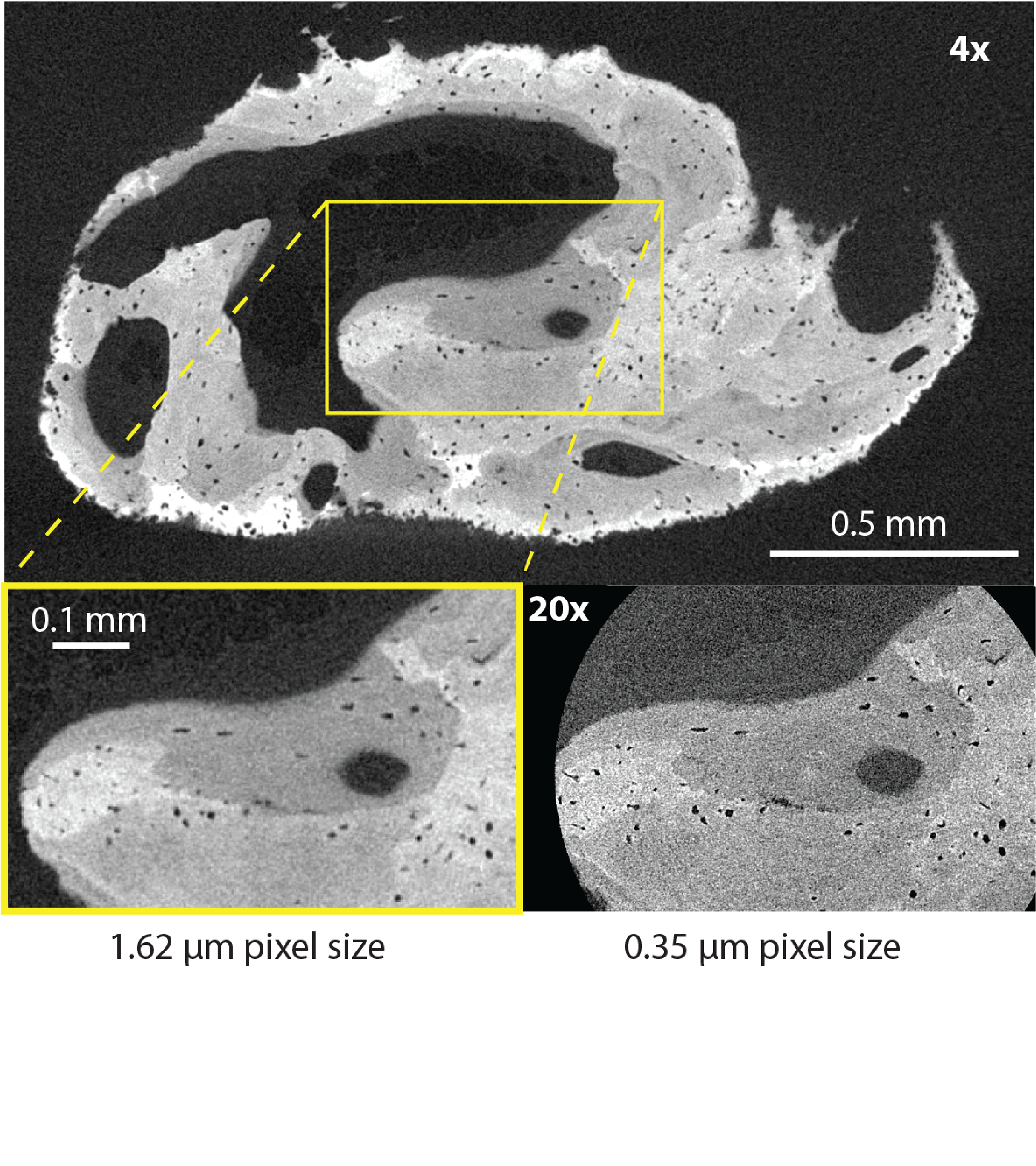
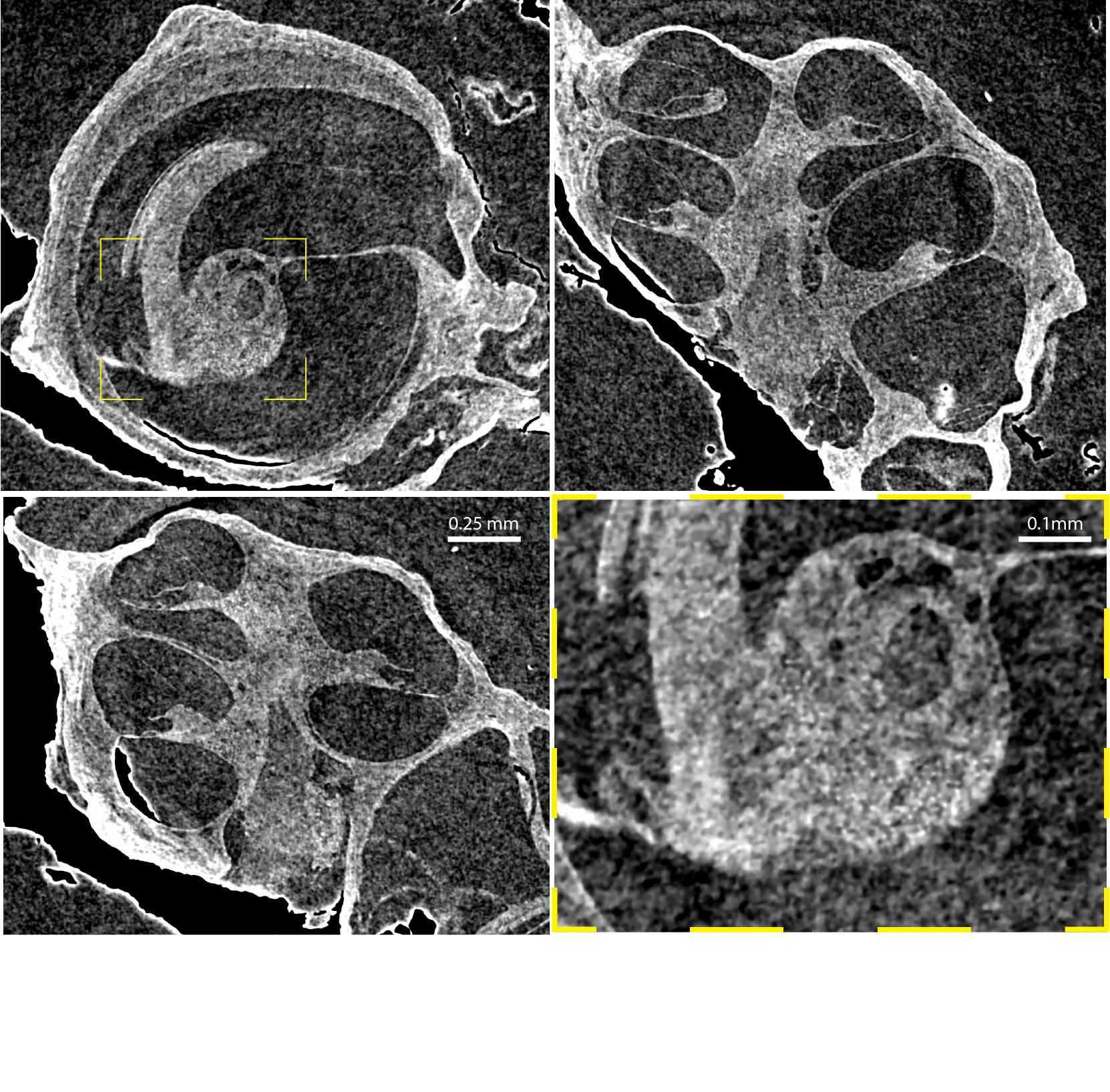
- smallest effective pixel size: 0.3 µm
- reconstructed volume after single scan: 3052 × 3052 × 2400 voxels
- reconstructed volume after 'off-axis' scan: 4624 × 4624 × 2400 voxels
- typical scanning time (depending on application): 5 - 480 min
X-ray detector
- temperature-stabilized high dynamic GE DXR
- 7 MP (3072 x 2400)
- 100 µm pixel size
Nanofocus X-ray source
- spot size: < 1 µm
- acceleration voltage: 40 - 180 kV
- maximal power: 20 W
- available targets: tungsten on CVD diamond & molybdenum
Scanning parameters
- maximal sample diameter: 80 mm
- maximal sample height: 70 mm
- maximal sample weight: 25 kg
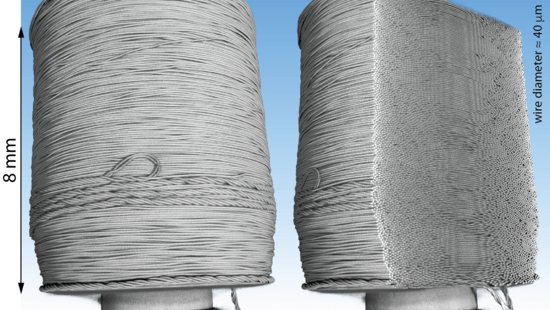
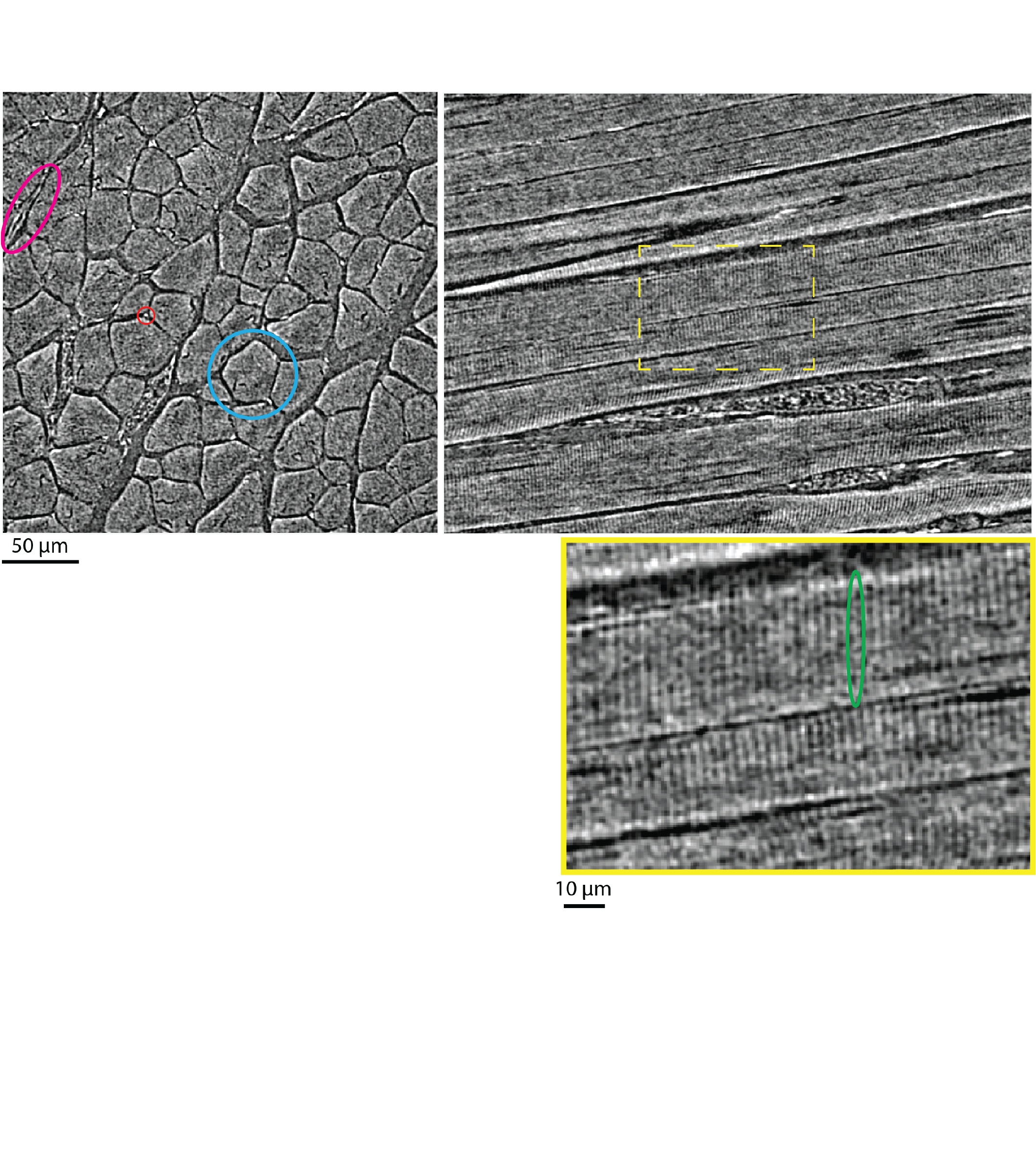
- smallest effective pixel size: 40 nm
- reconstructed volume after single scan: 1994 × 1994 × 2048 voxels
- reconstructed volume after 'off-axis' scan: 3756 × 3756 × 2048 voxels
- typical scanning time (depending on application): 1 - 24 h
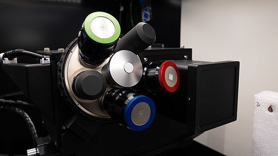
Detector system
- detector turret with multiple objectives at different magnifications
- each objective features optimized scintillator
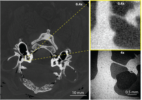
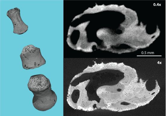
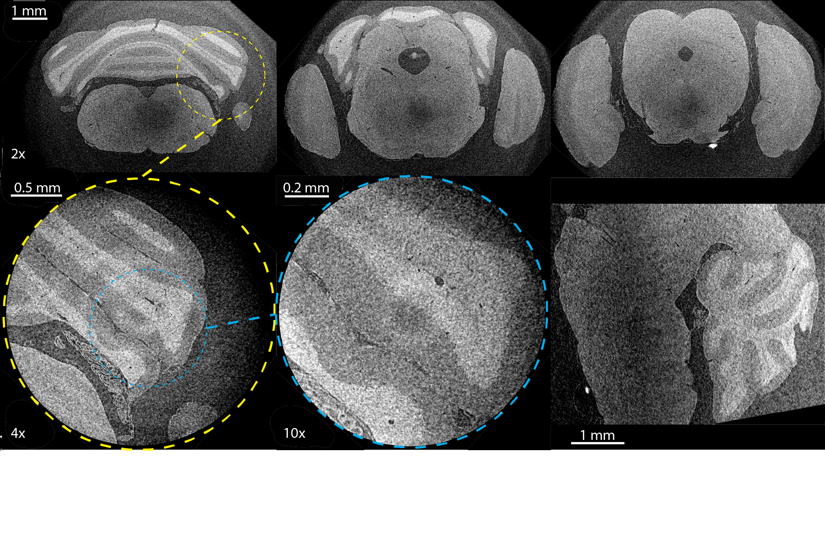
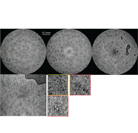
- available objectives: 0.4x, 4x, 20x, 40x
- 4 MP (2048 x 2048)
- 13.5 µm pixel size
Microfocus X-ray source
- spot size: < 5 µm
- acceleration voltage: 30 - 160 kV
- maximal power: 25 W

Scanning parameters
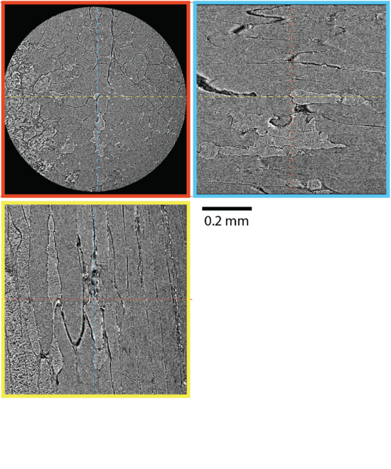
- maximum field of view: 6.5 mm
- maximum scannable height: 290 mm
- maximal sample weight: 6 kg
- smallest effective pixel size: 0.5 µm
- reconstructed volume after single scan: 2048 × 2048 × 2048 voxels
- reconstructed volume after 'off-axis' scan: 3700 × 3700 × 2048 voxels
- typical scanning time (depending on application): 1 - 24 h
Detector system
- detector turret with multiple objectives at different magnifications
- each objective features optimized scintillator
- available objectives: 2x, 4x, 10x
- 4 MP (2048 x 2048)
- 6.5 µm pixel size
Liquid metal X-ray source
- MetalJet D2+ (Excillum)
- jet material (target): I1 (68 % Ga, 22 % In, 10 % Sn)
- e-beam size (W x H): 40-80 µm x 10 µm
- spot size @60 µm e-beam width (W x H):
- @40 kV: 3.4 µm x 10.6 µm
- @70 kV: 7.2 µm x 12.2 µm
- acceleration voltage: 21 - 70 kV
- maximal power: 250 W
More information and any inquiries directly to Exciscope can be made through https://exciscope.com/contact/