Technologies
The Material Testing Service Unit combines state-of-the-art instrumentation including multimode plate readers, qPCR systems, fluorescence and electron microscopy, rheological characterization, and advanced fabrication technologies such as 3D printing and melt electrowriting enabling a comprehensive workflow from material fabrication and structural analysis to biological response assessment.
For more information, please refer to the instrument descriptions below or contact us directly.
Interested in our services?
Contact us for more details or a price estimation of our services.
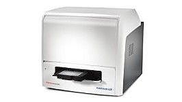
A high-performance, versatile microplate reader designed for bioscience applications, offering flexible detection modes and advanced data handling
- Multimode detection: Supports up to five measurement technologies: absorbance (UV-Vis), fluorescence intensity (incl. FRET), luminescence (direct/filtered), AlphaScreen/AlphaLISA and time-resolved fluorescence (TR-FRET/hTRF) for broad assay compatibility.
- Flexible measurement modes: Includes endpoint, kinetic, spectral, kinetic spectra and multipoint assays to suit diverse experimental designs.
- Smart optics and dynamic range: Combines filters and monochromators with automatic dynamic range selection to optimize signals across wells.
- Integrated features: Real-time spectral & kinetic displays, automatic calibration, intelligent safety controls and support for CO₂/O₂ gas control for cellbased assays.
- Broad plate compatibility & automation: Reads plates from 6 to 1536 wells and supports robotic integration.
- Certifications & validation: HTRF™ certified and Promega DLReady™ validated, ensuring compatibility with key assay formats.
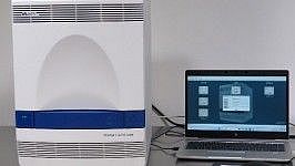
The Applied Biosystems 7500 Real Time PCR System is a reliable and versatile quantitative PCR platform for nucleic acid quantification, gene expression analysis, and multiplex PCR assays. It combines precise temperature control with sensitive fluorescence detection to deliver accurate, realtime amplification data for up to 96 samples per run.
- Real-time fluorescence detection: Optical system with a CCD camera and five excitation/emission filters enables monitoring of PCR products as they accumulate.
- Multiplexing: Can detect up to five distinct targets per well in a single reaction using multiple fluorophores.
- Wide dynamic range: Linear quantification across nine orders of magnitude, allowing detection from very low to high target amounts.
- High sensitivity: Capable of detecting single copy target molecules with high confidence.
- Flexible dye compatibility: Calibrated for common fluorescent chemistries including SYBR Green, FAM, VIC, ROX, and Cy5.
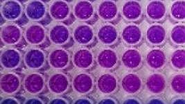
The MTT Assay using CellTiter Blue reagent is a fluorescence based method to assess cell viability and metabolic activity. Viable cells convert the non fluorescent resazurin dye into highly fluorescent resorufin through active cellular metabolism. The fluorescence signal is directly proportional to the number of living cells, making the assay suitable for cytotoxicity, proliferation, and drug screening studies.
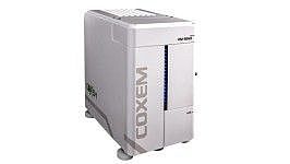
The Scanning Electron Microscope (SEM) uses a focused electron beam to generate high-resolution images of a sample’s surface morphology. It provides detailed three-dimensional-like visualization at the micro- and nanoscale by detecting electrons emitted from the sample surface.
Typical applications:
- Surface morphology and topography analysis
- Microstructure characterization
- Fiber and scaffold architecture imaging
- Particle size and shape analysis
- Quality control and failure analysis
Key features:
- High magnification and depth of field
- Micro- to nanoscale resolution
- Various imaging modes (secondary electrons, backscattered electrons)
- Elemental analysis possible when coupled with EDS (energy-dispersive spectroscopy).
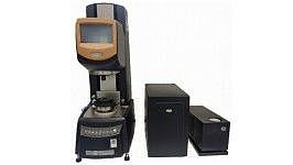
The TA Discovery HR-10 rheometer enables quantitative mechanical and rheological characterization of soft and hydrated materials under controlled conditions. It measures viscoelastic properties such as storage modulus (G′), loss modulus (G″), viscosity, yield stress, and stress–strain behaviour of hydrogels, bioinks, polymers, and soft tissues.
Typical applications:
- Stiffness comparison of biomaterials
- Gelation and crosslinking studies
- Frequency, amplitude, and strain sweep analysis
- Flow and viscosity measurements
- Stability assessment under physiologically relevant conditions
Key features:
- High sensitivity torque control for soft materials
- Oscillatory and rotational testing modes
- Temperature-controlled measurements
- Suitable for biomaterial development and rheological optimization.
The Axolotl 3D printer is designed for high-resolution extrusion-based fabrication of soft materials and bioinks. It can be used to produce customized 3D constructs, patterned hydrogels, and multi-material structures with controlled geometry and architecture.
This service supports rapid prototyping of scaffolds, organ-on-chip components, and test structures for mechanical, biological, or imaging studies, including reproducible fabrication of small-scale research models.
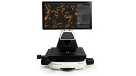
The EVOS 5000 microscope is an automated digital inverted fluorescence microscope used for routine cell imaging, including live/dead assays and immunofluorescence (IF). It allows fast imaging with multiple magnifications (4×, 10×, 20×, 60×) and different light channels.
Typical use:
- Live/Dead assays: observe cell morphology and viability using fluorescence dyes.
- Immunofluorescence (IF): detect specific proteins or markers labeled with fluorophores.
Light channels / filters:
- DAPI: blue channel (nuclei staining).
- FITC: green fluorescence.
- Red: red fluorophores (e.g., Texas Red).
- Trans (transmitted light): brightfield imaging.
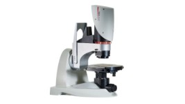
The LEICA DVM6 is a high-resolution digital microscope designed for detailed surface imaging and analysis. It combines optical zoom, automated settings, and large depth of field for fast documentation and measurement.
Typical use:
- Surface inspection and morphology analysis
- Material or sample characterization
- High-resolution imaging and documentation (not mainly for live cell fluorescence)
Magnification / optics:
- Wide zoom range (macro to micro scale depending on lens)
- Automatic calibration and focus
Imaging modes:
- Brightfield/reflected light
- Oblique and ring illumination for surface contrast
- Extended depth of field (EDF)
- 2D/3D surface visualization and measurement
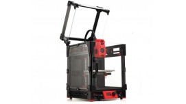
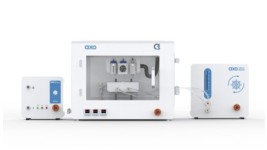
The MEW (Melt Electrowriting) printer, based on a Voron 2.0 platform, is a high-precision additive manufacturing system used for controlled deposition of microfibers and scaffold fabrication using electrically driven melt extrusion.
Typical use:
- Fabrication of microstructured scaffolds
- Tissue engineering and biomaterial research
- Precise fiber deposition and patterning
Technical parameters:
- Print head temperature: up to 250°C melting range (high-temperature printing capability)
- Nozzle: G27 / G25
- High voltage: up to 20 kV (electrostatic fiber drawing)
- Pressure controller: up to 2 mPa (controlled melt extrusion)
Key features:
- High positional accuracy (Voron 2.0 motion system)
- Fine fiber diameter control
- Stable electric field for MEW process
- Customizable parameters for reproducible scaffold structures.