LAser-assisted RObot-guided CArtilage REgeneration (LAROCARE)
Motivation
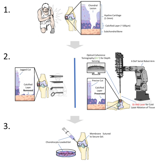
Joint injuries and cartilage degeneration are major health problems affecting millions of people worldwide, leading to pain, loss of mobility and reduced quality of life. Current treatments for cartilage defects are often invasive, lack precision and produce inconsistent results in cartilage regeneration. In response, the LAROCARE project aims to develop a minimally invasive, high-precision surgical solution that combines robotic technology with laser-based techniques to prepare defective cartilage tissue. This approach offers precise cutting capabilities, minimal collateral tissue damage and improved cartilage repair outcomes through robotic guidance and real-time feedback.
Project Overview and Partners
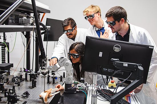
The LAROCARE project is an interdisciplinary collaboration to address the challenges of treating cartilage defects with advanced technology. The project integrates expertise from several fields:
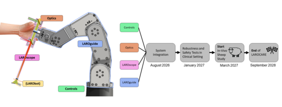
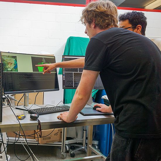
- Robotics and mechatronics – BIROMED-Lab, led by Prof. Georg Rauter: The team is developing the robotic components and control systems essential for this highprecision intervention.
- Laser and optical technologies – CIO, led by Prof. Ferda Canbaz: This team designs the laser systems to cut defective cartilage with minimal tissue damage, allowing precision unattainable by traditional manual methods. The system also includes an optical coherence tomography (OCT) fibre for intra-operative imaging and sensing.
- Cell Biology and Cartilage Engineering, Prof. Andrea Barbero: This team focuses on regenerative tissue engineering, where chondrocytes (cartilage cells) are encapsulated in biocompatible gels for improved tissue integration. In addition, their research focuses on replacing the usual cell source for growing a cartilage implant from the joints with the nasal region, which promises to be less invasive.
- Clinical Veterinary Expertise, Prof. Antonio Pozzi - Animal studies in sheep will provide valuable insight into the efficacy and safety of the laser-assisted robotic system.
- 3D imaging and microtomography, Dr. Georg Schulz - This team will ensure that structural and cellular imaging meets the project's requirements to assess cartilage regeneration at the microscopic level.
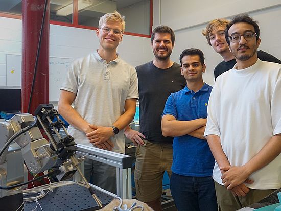
Project leader: Michael Sommerhalder and Jan Schimmelpfennig


Barbero Lab (University of Basel):
https://biomedizin.unibas.ch/en/research/research-groups/barbero-lab/
Center for Intelligent Optics (CIO) (University of Basel):
https://dbe.unibas.ch/en/research/laser-and-robotics/biomedical-laser-and-optics-group/
Small Animal Sugery Department (University of Zürich):
https://www.tierarzt-prof-antonio-pozzi.com/en/prof-antonio-pozzi.html
- Master Thesis: OCT-Based Adaptive Orientation Control (PDF, 4.10 MB)
- Master Thesis: Constraint-Compliant Control for Laser Ablation (PDF, 4.06 MB)
- Master Thesis: Markerless Preop-Registration with Stereovision (PDF, 4.04 MB)
- Internship: Mechatronic System Development (PDF, 4.04 MB)
- Master Thesis: Trinocular Endoscope Stereomatching (PDF, 4.06 MB)

