Detecting Early Choroidal Changes Using Piecewise Rigid Image Registration and Eye-Shape Adherent Regularization
This project takes place in the frame of an international cooperation between the University of Applied Sciences of Biel (project center), the Department of Biomedical Engineering of the University of Basel (academic supervision), Department of Ophthalmology (clinical support), Guangzhou and Hong Kong (clinical partners). The research is focused on the development of an automatic algorithm for the detection of subtle changes in the area of the human choroid, a soft tissue located in the posterior part of the eye between the retina and sclera.
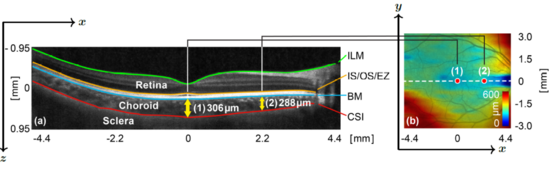
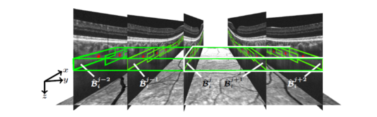
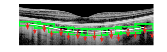
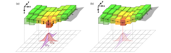
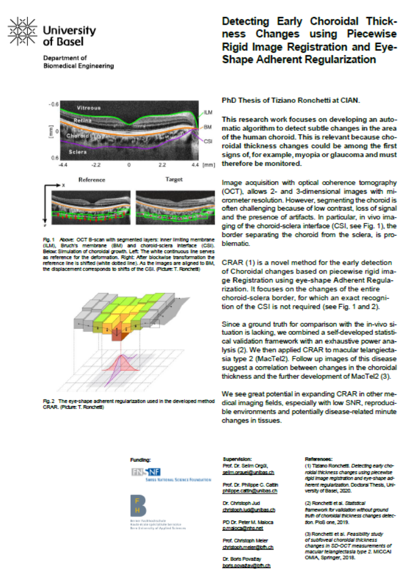
Significant modifications in the choroidal thickness are used in clinical applications as indicator of ocular disorders (e.g. myopia) up to diseases (glaucoma). Recognizing temporal changes in the thickness of the choroid and retina at an early stage is a crucial factor in the prevention and treatment of such visual impairments. The thickness of the choroid is the distance between the Bruch’s Membrane (BM) and the Choroid-Sclera Interface (CSI), see Fig. 1. Choroidal maps visualize the locally varying thickness of the choroid and can aid to extract essential clinical information, see Fig. 1b. In the developed method, “Detecting early Choroidal changes using piecewise rigid image Registration and eye-shape Adherent Regularization” (CRAR), the volume around the CSI is subdivided in partially overlapping cuboidal subregions (see Fig. 2), utilizing the rigid BM as a reference surface. Using a multiresolution approach, a 3D regularized block-matching registration of the CSI is conducted. As the images are aligned to the rather rigid BM, the displacement corresponds to shifts of the CSI interface. Thus, it is possible to determine the displacement field around the CSI layer and to use its outcome to quantify choroidal growth, see Fig 3. The position of the mismatched block (red) in the 3D registration is corrected by regularization, see Fig. 4(a). This has a smoothing effect, because the directly neighboring blocks (yellow) counteract this movement. Distant blocks (light green) have weaker influence. The mismatched blocks are not individually corrected. Instead, the entire neighborhood is moved until the block configuration with the least bending energy is reached, see Fig. 4(b). In order to overcome the problem of the absence of a ground truth for comparison with the in vivo situation, we developed a statistical validation framework for automated choroidal thickness changes detection, in which a method purely based on the common agreement between the algorithm and all experts is combined with an exhaustive power analysis approach. In a retrospective study in collaboration with the Moorsfield Eye Hospital in London, we further applied CRAR on macular telangiectasia type 2 (MacTel2). The analysis of follow up images of this disease suggests that there might be a correlation between changes in the choroidal thickness and the further development of MacTel2.
The further refinement of the presented method CRAR can provide an objective and sensitive tool to analyze and monitor the progress of myopia, and beyond. We also see great potential in expanding CRAR in other medical imaging fields (e.g. OCT-dermatology), especially where low signal to noise and reproducible environments can be found, which makes a reliable segmentation of tissues very challenging, and where minute changes in tissues have to be monitored as potentially disease-related.
Project leader: Tiziano Ronchetti


T. Ronchetti, C. Jud, P. M. Maloca, S. Orgül, A. T. Giger, C. Meier, H. P. Scholl, R. K. M. Chun, Q. Liu, C.-H. To, B. Považay, and P. C. Cattin, “Statistical framework for validation without ground truth of choroidal thickness changes detection”, PloS one, vol. 14, no. 6, p. e0218776, 2019. read
T. Ronchetti, P. Maloca, E. R. de Carvalho, T. F. Heeren, K. Balaskas, A. Tufail, C. Egan, M. Okada, S. Orgül, C. Jud, and P. C. Cattin, “Feasibility study of subfoveal choroidal thickness changes in spectral-domain optical coherence tomography measurements of macular telangiectasia type 2”, in Computational Pathology and Ophthalmic Medical Image Analysis. Springer, 2018, pp. 303–309. read
T. Ronchetti, P. Maloca, C. Jud, C. Meier, S. Orgül, H. P. Scholl, B. Považay, and P. C. Cattin, “Detecting early choroidal changes using piecewise rigid image registration and eye-shape adherent regularization”, in Fetal, Infant and OphthalmicMedical Image Analysis. Springer, 2017, pp. 92–100. read
T. Ronchetti, P. Maloca, C. Meier, S. Orgül, C. Jud, P. Hasler, B. Považay, and P. C. Cattin, “Intensity-based choroidal registration using regularized block matching”, in Proceedings of the Ophthalmic Medical Image Analysis Third International Workshop (OMIA), 2016, pp. 33–40. read
No open projects currently