Diffusion Models for Contrast Harmonization of Magnetic Resonance Images
Magnetic resonance (MR) images obtained from various sources frequently exhibit variations in image contrast due to differences in acquisition parameters or the type of scanner employed. In long-term studies, maintaining longitudinal comparability is crucial; however, the aforementioned contrast differences can compromise this comparability, resulting in biased outcomes when utilizing automated evaluation tools. This study introduces a diffusion model-based method for harmonizing contrast.
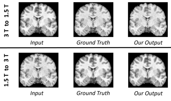
We use a dataset of scans from Multiple Sclerosis patients and healthy controls, with each participant scanned in two MR scanners at different magnetic field strengths (1.5 T and 3 T). This results in a paired dataset that highlights scanner-specific, and thus field strength-specific, differences. We use a diffusion model to map slice-by-slice from source to target contrast, in both directions: from 3 T to 1.5 T and 1.5 T to 3 T. To change only the contrast while preserving anatomical information, we use the original image to guide the translation process. The goal is for the transformed scans to display enhanced comparability with scans of the target contrast for subsequent tasks. Therefore, in addition to mean squared error and histogram analysis, we evaluate the method's effectiveness by segmenting cerebrospinal fluid, grey matter, and white matter, before and after contrast harmonization. We achieve good and consistent results in both mapping directions. More details can be found in (1).
Project leader: Alicia Durrer
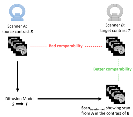
Scans acquired with different MR scanners show differences in image contrast. This can impair longitudinal comparability, e.g. in the case of monitoring Multiple Sclerosis progression. The goal of this project is to transform scans from source to target contrast to ensure comparability between different scans.