Improving Cranial Navigation with an Advanced Microscope
Neuronavigation has become an essential tool in cranial and spinal neurosurgery. Currently it is based on preoperatively acquired images of modalities like CT or MRI, which are then co-registered with anatomic landmarks on the patient's skin or using surface matching via laser scanning.
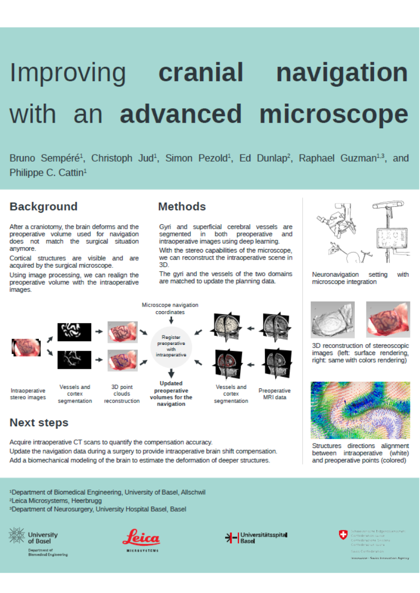
The major biological factor for accuracy loss in cranial navigation is the so called brain shift. It occurs after a craniotomy, when the brain starts to deform in the cranial cavity, and therefore mismatches the preoperative volumes. This remains one of the most significant shortcomings of neuronavigation that can so far only be corrected using an additional intraoperative imaging such as ultrasound, CT or MRI.
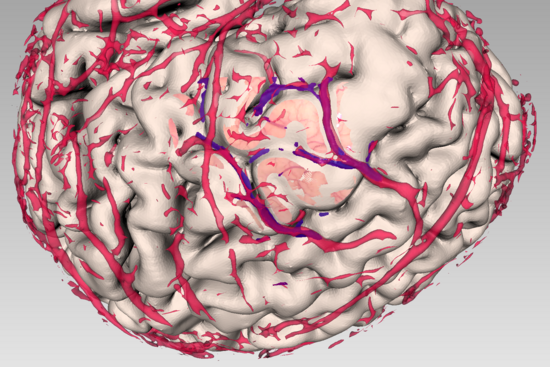
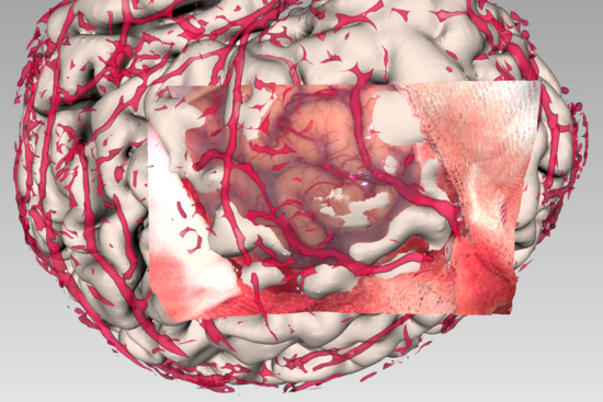
In contrast to these methods, this project aims to compensate this deformation by relying on an advanced surgical microscope, already present in the operating room. During cerebral surgeries, a portion of the cerebral cortex is exposed, exhibiting the unique pattern of gyri, sulci and superficial vessels. These structures are acquired in the preoperative MR volumes and by the surgical microscope during the surgery.
By leveraging the stereoscopic capabilities of the new advanced microscopes of Leica Microsystems, it is possible to reconstruct the intraoperative site in three dimensions. Combining these advancements with the new developments in artificial intelligence, one can extract the pattern of the brain surface both preoperatively and intraoperatively, and use this information to update the navigation volumes to match the actual surgical context.
The project was funded by the Swiss Innovation Agency Innosuisse and is carried out in collaboration between the Center of medical Image Analysis & Navigation (CIAN) of the Department of Biomedical Engineering, University of Basel, the Neurosurgery Department at University Hospital Basel (USB), and Leica Microsystems (Switzerland) Ltd. (Heerbrugg, SG).
Project leader: Christoph Jud


