Dynamic and interventional MRI

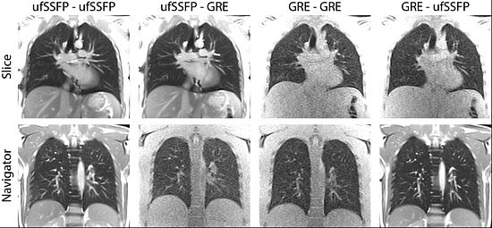
Capturing motion with MRI can be wanted or unwanted. Typically, abdominal or chest motion is a major concern in the treatment of tumors using MR-guided high-intensity focused ultrasound (MRgHIFU) or proton therapy. We are active in the field of MR-guided interventions since more than a decade and provide dedicated methods for tracking organ motion to our national and international partners.
Project leader and contact
Project members
Collaborators
- Prof. Dr. Tony Lomax – Paul Scherrer Institute
- Prof. Dr. Rares Salomir – HUG Geneva
- Prof. Dr. Philippe Cattin - University of Basel
Selected publications
Bauman, G., Lee, N. G., Tian, Y., Bieri, O., & Nayak, K. S. (2023). Submillimeter lung MRI at 0.55 T using balanced steady-state free precession with half-radial dual-echo readout (bSTAR). Magnetic Resonance in Medicine. https://doi.org/10.1002/mrm.29757
Bieri, O., Pusterla, O., & Bauman, G. (2022). Free-breathing half-radial dual-echo balanced steady-state free precession thoracic imaging with wobbling Archimedean spiral pole trajectories. Zeitschrift Fur Medizinische Physik, S0939-3889(22)00003-4. https://doi.org/10.1016/j.zemedi.2022.01.003
Breit, H.-C., & Bauman, G. (2022). Morphologic and Functional Assessment of Sarcoidosis Using Low-Field MRI. Radiology, 211760. https://doi.org/10.1148/radiol.211760
Pusterla, O., Heule, R., Santini, F., Weikert, T., Willers, C., Andermatt, S., Sandkühler, R., Nyilas, S., Latzin, P., Bieri, O., & Bauman, G. (2022). MRI lung lobe segmentation in pediatric cystic fibrosis patients using a recurrent neural network trained with publicly accessible CT datasets. Magnetic Resonance in Medicine. https://doi.org/10.1002/mrm.29184
Bauman, G., & Bieri, O. (2020). Balanced steady‐state free precession thoracic imaging with half‐radial dual‐echo readout on smoothly interleaved archimedean spirals. Magnetic Resonance in Medicine, 84(1), 237–246. https://doi.org/10.1002/mrm.28119
Bauman, G., Pusterla, O., Santini, F., & Bieri, O. (2018). Dynamic and steady-state oxygen-dependent lung relaxometry using inversion recovery ultra-fast steady-state free precession imaging at 1.5 T: Oxygen-Dependent Lung Relaxometry. Magnetic Resonance in Medicine, 79(2), 839–845. https://doi.org/10.1002/mrm.26739
Pusterla, O., Sommer, G., Santini, F., Wiese, M., Lardinois, D., Tamm, M., Bremerich, J., Bauman, G., & Bieri, O. (2018). Signal enhancement ratio imaging of the lung parenchyma with ultra-fast steady-state free precession MRI at 1.5T: SER Imaging of the Lung Parenchyma. Journal of Magnetic Resonance Imaging, 48(1), 48–57. https://doi.org/10.1002/jmri.25928
Bauman, G., & Bieri, O. (2017). Matrix pencil decomposition of time‐resolved proton MRI for robust and improved assessment of pulmonary ventilation and perfusion. Magnetic Resonance in Medicine, 77(1), 336–342. https://doi.org/10.1002/mrm.26096
Nyilas, S., Bauman, G., Sommer, G., Stranzinger, E., Pusterla, O., Frey, U., Korten, I., Singer, F., Casaulta, C., Bieri, O., & Latzin, P. (2017). Novel magnetic resonance technique for functional imaging of cystic fibrosis lung disease. European Respiratory Journal, 50(6), 1701464. https://doi.org/10.1183/13993003.01464-2017
Pusterla, O., Bauman, G., Wielpütz, M. O., Nyilas, S., Latzin, P., Heussel, C. P., & Bieri, O. (2017). Rapid 3D in vivo 1H human lung respiratory imaging at 1.5 T using ultra-fast balanced steady-state free precession: Ultra-fast SSFP for 3D Lung Functional MRI. Magnetic Resonance in Medicine, 78(3), 1059–1069. https://doi.org/10.1002/mrm.26503
