Neuro MRI


Brain
From a physicist’s perspective the brain represents the ideal playground for MRI physics and methods development: it is not too big, there is no bulk motion, it is near spherical (for physicists only), it has a rich and complex microstructure to explore, it is well characterized, and so on. Or to put it simple: if some of your development does not work in the brain, it does not work anywhere else. As a result, imaging of the brain is, apart from phantoms, our most preferable in-vivo test object.
Spinal cord
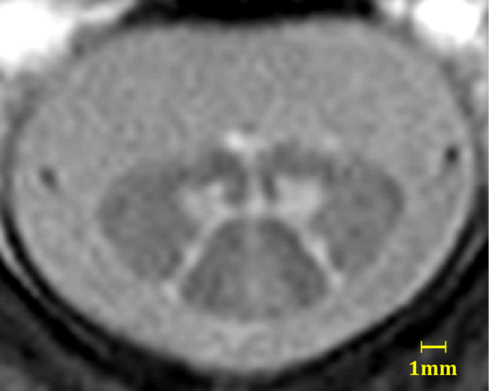
The spinal cord, a long and thin nervous tissue of tubular shape, extends from the brain stem to the lumbar vertebrae and MRI faces increasing challenges along this route. Generally, MRI of the spinal cord typically requires high resolutions (the typical diameter is only 7 – 13mm), a large field-of-view, and is complicated by physiologic chest motion for the thoracic and lumbar segments. The gray matter extends from the brain into the spinal cord but changes from the outside to the inside appearing in cross-sections with a shape that resembles a butterfly. And this is where the problem lies: capture the butterfly with MRI.
Our in-house developed method, termed AMIRA, was solely designed to fulfill this purpose;, it offers superior white-gray matter contrast to resolve the butterfly and is currently applied in a range of clinical studies, such as spinal muscular atrophy (SMA) and multiple sclerosis (MS). Besides AMIRA, we also investigate common contrast mechanisms, such as diffusion, for imaging the spinal cord.
Project leader and contact
Project members
Collaborators
- Dr. med. Regina Schläger
- Dr. med. Janina Wendenburg
- Dr. med. Eva Kesenheimer
- Prof. Dr. med. Cristina Granziera