Musculoskeletal MRI
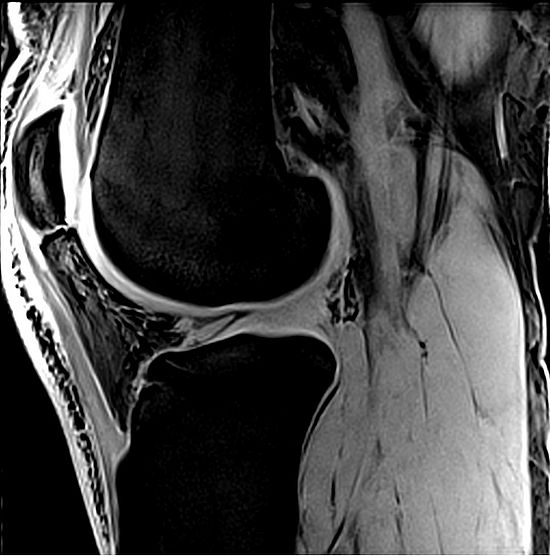
We work on the development of quantitative methods for musculoskeletal MRI.
We have developed methods for imaging cartilage and tendons at high and ultra high field strengths, and we are currently focusing on muscle MRI, especially quantitative and dynamic imaging.
Please visit the dedicated group page of BaMM (Basel Muscle MRI) for more information!
Project leader and contact
Project members
Collaborators
- Prof. Reinald Brunner - Universitätskinderspital Basel
- Dr. Nicolas Place - University of Lausanne
- Prof. Anna Pichiecchio - Fondazione Mondino, Pavia, Italy
- Dr. Giorgio Tasca - Policlico Gemelli, Rome, Italy
- Prof. Fengdan Wang - Peking Union Medical College, Beijing, China
- Esther Steijvers - Scannexus, Maastricht, The Netherlands
- Prof. Dr. Siegfried Trattnig - Medizinische Universität Wien, Austria
- Dr Vladimir Juras - Medizinische Universität Wien, Austria
- Prof Miika Nieminen - University of Oulu, Finland
- Dr. Victor Casula - University of Oulu, Finland
Selected publications
Felisaz, P. F., Belatti, E., Deligianni, X., Bergsland, N., Santini, F., Paoletti, M., Solazzo, F., Germani, G., Cortese, A., Vegezzi, E., Bieri, O., Bastianello, S., & Pichiecchio, A. (2020). Variable echo time imaging for detecting the short T2* components of the sciatic nerve: a validation study. Magnetic Resonance Materials in Physics, Biology and Medicine. https://doi.org/10.1007/s10334-020-00886-w
Zbýn, Š., Schreiner, M., Juras, V., Mlynarik, V., Szomolanyi, P., Laurent, D., Scotti, C., Haber, H., Deligianni, X., Bieri, O., Nieminen, M. T., & Trattnig, S. (2020). Assessment of Low-Grade Focal Cartilage Lesions in the Knee With Sodium MRI at 7 T: Reproducibility and Short-Term, 6-Month Follow-up Data. Investigative Radiology, 55(7), 430–437. https://doi.org/10.1097/RLI.0000000000000652
Bachmann, E., Rosskopf, A. B., Götschi, T., Klarhöfer, M., Deligianni, X., Hilbe, M., Pfirrmann, C. W. A., Snedeker, J. G., & Fischer, M. A. (2019). T1- and T2*-Mapping for Assessment of Tendon Tissue Biophysical Properties: A Phantom MRI Study. Investigative Radiology, 54(4), 212–220. https://doi.org/10.1097/RLI.0000000000000532
Hager, B., Walzer, S. M., Deligianni, X., Bieri, O., Berg, A., Schreiner, M. M., Zalaudek, M., Windhager, R., Trattnig, S., & Juras, V. (2019). Orientation dependence and decay characteristics of T2* relaxation in the human meniscus studied with 7 Tesla MR microscopy and compared to histology. Magnetic Resonance in Medicine, 81(2), 921–933. https://doi.org/10.1002/mrm.27443
Latta, P., Juras, V., Kojan, M., Starcuk, Z., Deligianni, X., Bieri, O., Szomolanyi, P., Frollo, I., & Trattnig, S. (2019). The Experimental Setup for T-2* Mapping in Achilles tendon and Enthesis. In J. Manka, J. Svehlikova, V. Witkovsky, & I. Frollo (Eds.), 2019 Proceedings of the 12th International Conference on Measurement (measurement 2019) (pp. 141–144).
Hornakova, L., Juras, V., Kubovy, P., Hadraba, D., Gerych, D., Stursa, P., Deligianni, X., Bieri, O., Trattnig, S., & Jelen, K. (2018). In vivo assessment of time dependent changes of T2* in medial meniscus under loading at 3T: A preliminary study. Journal of Applied Biomedicine, 16(2), 138–144. https://doi.org/10.1016/j.jab.2017.12.001